Diagnosing CRGV

In dogs presented with possible CRGV (on the basis of skin lesion appearance), consideration should be given to baseline measurement of at least serum creatinine, along with a platelet count and, ideally, urinalysis. Glomerular filtration rate and/or symmetric dimethylarginine can be assessed in non-azotemic cases in an attempt to identify early reduction in glomerular filtration rate. In suspected cases, bloodwork and urinalysis should be reassessed within 24 to 48 hours, and the owner should be asked to subjectively monitor urine output.
Histopathology
Histopathologically, dogs with CRGV have a thrombotic microangiopathy (TMA), which is characterised by inflammation and damage to vascular endothelium, leading to platelet activation and aggregation. This results in widespread formation of microthrombi. Formation of microthrombi leads to consumptive thrombocytopenia and can result in occlusion of the vessel lumen, causing ischemic damage and death of affected tissue. Thrombotic microangiopathy in CRGV seems to primarily target the skin and kidneys.
Skin Lesion Appearance
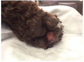
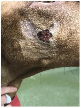
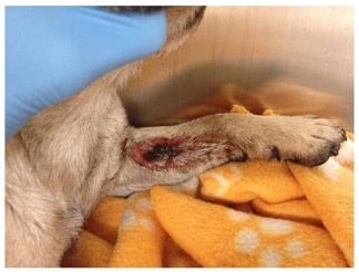
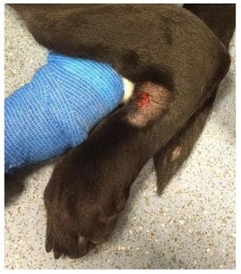
Patient presentation is commonly due to skin lesions that range from small, superficial abrasions (0.5 cm) to large areas of full-thickness ulceration and necrosis (>30 cm) with surrounding erythema and oedema (Figures 1-4). The average time from development of skin lesions to the development of acute kidney injury (AKI) is 3 days. In one study, most dogs (80.9%) presented with a skin lesion on a limb, shoulder or digit. Of the 144 dogs with limb lesions, 106 had lesions affecting the pads, foot, or digits (73.6%). Lesions of the body or an unspecified location were present in 20.8% of dogs. Lesions were present in the oral cavity in 8.4% of dogs. In only 1.1% of cases, AKI was documented prior to development of skin lesions. A single lesion was observed in 61.2% of dogs, and 69/178 dogs (38.8%) had multiple lesions.
Some dogs present with lameness, with the pet owner not identifying a skin lesion(s). A proportion of dogs remain clinically well and recover from skin lesions uneventfully without developing detectable AKI. In one study, 21.7% of dogs in contact with a dog with CRGV were reported to develop skin lesions without developing AKI. Clinical signs in dogs that develop AKI can include vomiting, diarrhoea, polyuria/polydipsia, oliguria, anuria, lethargy, anorexia, hypothermia, icterus, and petechiae. Neurologic signs were identified in 18.6% of dogs during illness and in 1.1% at presentation.
Antemortem Diagnosis
Antemortem diagnosis can be challenging because there is no single non-invasive diagnostic test available. A combination of skin lesions and AKI ± thrombocytopenia and/or hyperbilirubinaemia are most likely to help reach a diagnosis of CRGV. Confirmation of disease requires post-mortem examination of renal tissue.
Renal histopathology is the gold standard for identification of the thrombotic microangiopathy process therefore a diagnosis of azotemic CRGV is commonly achieved post-mortem, as renal biopsy is rarely recommended in patients with AKI.
The most common hematologic abnormality when a patient is presented with AKI is thrombocytopenia which is present in nearly 84% of patients; anaemia is present in 29%. Blood smear examination may identify evidence of microangiopathic hemolysis (i.e., Burr cells, acanthocytes, schistocytes).
The most common biochemical abnormalities at presentation are elevated serum urea concentration in (91% of cases), elevated serum creatinine concentration (86%) and elevated serum liver enzyme activities (72%). Hyperphosphatemia is seen in just over 82% of patients and hyperbilirubinaemia in just over half. Over 40% of dogs on presentation have reduced or absent urine production.

FIGURE 1 Well-demarcated, erosive lesion on the digital pad of a crossbreed dog with CRGV

FIGURE 2 Well-demarcated, circular, ulcerative lesion on the face of a Hungarian vizsla with CRGV

FIGURE 3 Extensive, ulcerative, partially necrotic lesion over the medial antebrachium on a pug with CRGV

FIGURE 4 Well-demarcated, ulcerative lesion distal to the left tarsus on a Labrador retriever with CRGV
